
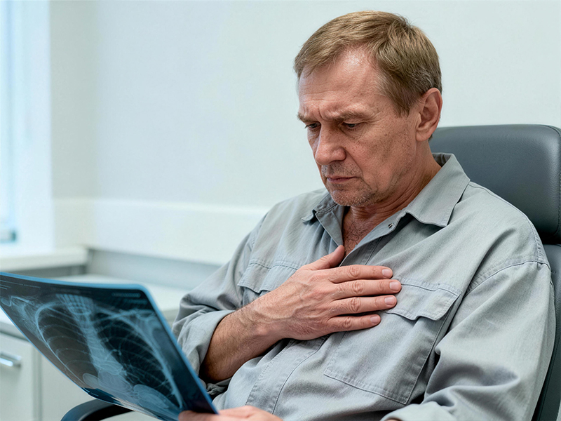
Patient records
Name: Ahmed Al-Rashid
Age: 45
Nationality: United Arab Emirates (Dubai)
Diagnosis: Traumatic chest deformity complicated by chronic empyema following a severe car accident (defect of large segments of the left 4th-8th ribs, fibrous lamination, and cardiac displacement).
Treatment Hospital: West China Hospital of Sichuan University (Department of Thoracic Surgery)
Ahmed posted a photo on his social media, looking ramrod straight in a tailored suit he had custom-made for his wedding, with the caption: "Thank you, China, for giving me a new suit of armor." Looking at this photo, my thoughts were instantly pulled back to a year ago, to the first time he walked into our office—his left side of his chest was severely sunken, like a flattened soda can, and even breathing was a luxury.
Ahmed survived a serious car accident, but was left with devastating aftereffects. Multiple rib fractures caused severe collapse of the left chest wall, leading to chronic empyema due to secondary infection. His left lung was tightly bound by a thick fibrous plate, unable to expand, and his entire left chest was severely deformed, with even his heart pushed to the right. He consulted doctors in many countries, only to be told that "the infection can be cleared, but the chest deformity cannot be reversed."
Faced with this devastating injury, which combined infection, deformity, and loss of function, our case manager turned to the Department of Thoracic Surgery at West China Hospital of Sichuan University. This department represents the pinnacle of expertise in China for treating extremely severe chest trauma and chest wall reconstruction. To help Ahmed understand this complex surgery, we coordinated with the expert team at West China Hospital to create a 1:1 three-dimensional reconstruction of his thoracic cavity using computer-aided design (CAD) technology. A 3D-printed model was then used to visually demonstrate the surgical plan: First, a pleurodesis was performed, peeling away the fibrous plaque binding the lung like peeling an orange; second, necrotic and infected rib segments were removed; and third, a 3D-printed, personalized titanium alloy chest wall prosthesis was implanted.
The surgery was performed in two stages. In the first stage, thorough debridement was conducted to remove the abscess and fibrous tissue, allowing his left lung, which had been compressed for over two years, to re-expand. Post-surgery, our case manager visited him daily in his ward, encouraging him to perform rigorous respiratory exercises. Six weeks later, the infection was completely under control, and the second stage surgery began. The doctors precisely implanted an "artificial rib"—designed to fit the mirror image of his healthy side of the chest and made of titanium alloy powder sintered by laser. This lightweight yet sturdy "armor" perfectly conformed to his anatomical structure, instantly restoring the symmetry of his chest.
Post-surgery, Ahmed's chest appearance returned to normal, and his lung function improved from 30% to 70%. After returning home, we arranged a year-long rehabilitation follow-up program for him, using regular lung function monitoring and 3D surface scans to assess the long-term stability of the implant. Now, wearing that sharp suit, he embarks on a new chapter in his life. This is not just a reconstruction of his chest, but a rebirth of his dignity and confidence.